

Capsule endoscopy of the small intestine: everything you would like to know
What is a small bowel capsule endoscopy:
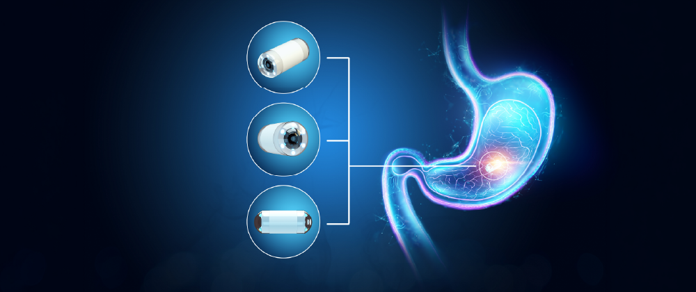
A capsule endoscopy is a non-invasive diagnostic test of your digestive system. The patient swallows a capsule no larger than a multivitamin, which contains a tiny camera, a transmitter, and a light.
As it passes through the stomach, small intestine, colon, and rectum, the capsule takes thousands of pictures over a period of 10-12 hours and transmits them to a recording device that the patient wears outside their clothes. After the few seconds it takes to swallow it, and once it enters the stomach, the patient does not feel it at all.
The examination can be performed on an inpatient basis, where necessary, but also on an outpatient basis, since a hospital stay is not necessary.
To which patients is the examination addressed?
The cases in which your doctor may recommend the examination are the following:
A) Investigating the cause of bleeding in the small intestine, usually in patients whose gastroscopy and colonoscopy were normal. This is the most common reason to perform a capsule endoscopy. Also, in patients with iron deficiency anemia without obvious digestive bleeding.
B) Diagnosis and subsequent monitoring of inflammatory bowel diseases. Capsule endoscopy can identify irritated and inflamed areas in the small intestine in diseases such as Crohn’s disease. It is often repeated during treatment to help us evaluate how well our patient is responding to it.
C) Diagnosis of small intestine cancer. Capsule endoscopy can show tumors in the small intestine that are not visible with classic gastroscopy and colonoscopy.
D) Diagnosis and monitoring of celiac disease. Capsule endoscopy is sometimes used to diagnose and monitor this disease, which is caused by an immune reaction to gluten. It is also used for other conditions related to malabsorption.
E) Evaluation for polyps in the small intestine. Certain familial genetic syndromes can cause polyps in the small intestine. A capsule endoscopy can check for these polyps.
F) Further evaluation after another imaging test, such as a CT scan. Often, imaging methods such as a CT scan or MRI scan give us unclear images or findings in the small intestine. In these cases, a capsule endoscopy can clarify these findings.
What preparation is needed?
The patient must have fasted since the previous afternoon. Often the doctor may choose to prescribe some preparation, which in any case is milder than that of a colonoscopy. After swallowing the capsule, the patient remains fasting for about 6 hours. Water intake is not restricted, neither before nor during the examination.
How is the diagnosis made?
The capsule passes through the digestive system, without needing to be regurgitated. The images it captures are transmitted wirelessly to a recording device that the patient wears outside his clothes.
The doctor uploads the data from the recording device using a system that combines images into video. This video is examined by the doctor for any pathologies of the small intestine.
How safe is the examination?
The capsule endoscopy is a safe examination with an extremely low rate of complications. Rarely, a capsule can get stuck in the digestive tract, mainly in patients who suffer from a narrowing of the digestive tract due to, for example, a tumor, Crohn’s disease or surgery in the area. In case the patient reports symptoms such as pain, bloating or vomiting, the possibility of intestinal obstruction can be ruled out with a CT scan.
According to the international literature, the incidence of complications is less than 1 in every 400 patients.
Dr. Evangelos Roussos regularly performs capsule endoscopy examinations at the Athens Medical Center, as well as at the Imperial College Healthcare Trust University Hospital in London.
Author


